Fluorescein angiography

Fluorescein angiography (FA)
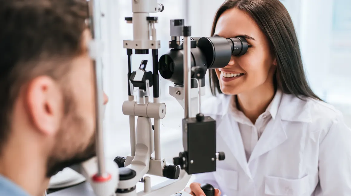
Fluorescein angiography (FA) is a medical imaging test used to evaluate the blood vessels in the retina and choroid, the light-sensitive tissues at the back of the eye. It involves injecting a fluorescent dye (fluorescein) into a vein in the arm, which then travels to the eyes and highlights the blood vessels.
FA is used to:
- 1. Diagnose and monitor eye conditions, such as:
– Diabetic retinopathy
– Age-related macular degeneration (AMD)
– Retinal vein occlusion
– Retinal artery occlusion - Detect and monitor treatment of:
– Retinal neovascularization (abnormal new blood vessel growth)
– Macular edema (fluid buildup in the macula) - Guide laser treatment and surgical interventions
The test involves:
- Injecting fluorescein dye into a vein in the arm
- Taking a series of photographs with a specialized camera
- Analyzing the images to evaluate blood vessel health and detect any abnormalities
FA is typically performed by an eye care professional (ophthalmologist or optometrist) and takes around 30-60 minutes to complete.
Note: While fluorescein angiography is not directly related to dentistry, it is an important diagnostic tool for eye health, and dentists often work closely with eye care professionals to ensure overall patient well-being.


