Optical coherence tomography

Optical Coherence Tomography (OCT)
Optical Coherence Tomography (OCT) is a non-invasive medical imaging technique that uses low-coherence interferometry to capture high-resolution, cross-sectional images of the internal structures of the eye. It is commonly used to:
- 1. Diagnose and monitor retinal conditions, such as:
– Macular degeneration
– Diabetic retinopathy
– Retinal detachment - Evaluate the optic nerve and detect conditions like glaucoma
- Monitor the progression of eye diseases and response to treatment
- Guide retinal surgery and other treatments
OCT works by:
- Splitting a low-intensity laser beam into two paths
- One path illuminates the eye, while the other serves as a reference
- Measuring the interference pattern created by the backscattered light
- Generating a detailed image of the eye’s internal structures
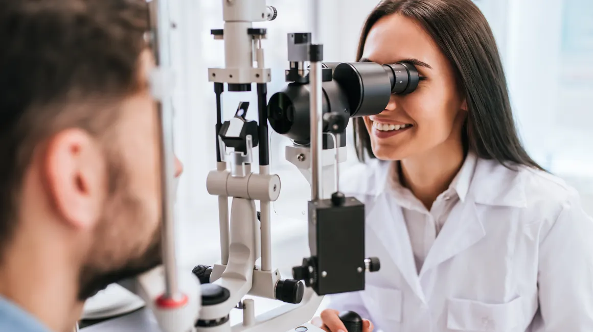
OCT is a painless and quick procedure, taking around 5-10 minutes per eye. It is performed by an eye care professional (optometrist or ophthalmologist) and is an essential tool for diagnosing and managing various eye conditions.
Note: While OCT is not directly related to dentistry, it is an important diagnostic tool for eye health, and dentists often work closely with eye care professionals to ensure overall patient well-being.


